













































































Are you trying to understand how photosynthesis actually works inside the chloroplast? This article offers you a chloroplast photosynthesis diagram to help you visualize every structure and every step of the process. Together, let's make this plant biology lesson accessible and fascinating!
General structure of the chloroplast and its components
The chloroplast is an extraordinary organelle where all the magic of plant life begins. Picture a true microscopic biological factory, measuring 7 to 8 µm long, protected by a double membrane and housing an aqueous space called the stroma. It's in this very stroma that you'll find the key enzymes enabling the production of organic matter through photosynthesis.

Membrane envelope and internal compartments
Each chloroplast is surrounded by two distinct membranes: a highly permeable outer membrane and a much more selective inner membrane. Between these two barriers lies a thin intermembrane space. This double membrane encloses the stroma, a gel-like aqueous fluid rich in active molecules.
- Outer membrane: Highly permeable, it lets through most molecules up to 5000 daltons, facilitating exchanges with the cytosol of the plant cell.
- Inner membrane: It strictly regulates what enters and leaves the stroma, ensuring a stable environment for chemical reactions.
- Intermembrane space: This thin gap between the two membranes facilitates the transfer of molecules and contributes to the overall structure.
- Chloroplast envelope: This double membrane, similar to that of mitochondria, reflects the endosymbiotic origin of these fascinating organelles.
Observed under a light microscope on a spinach leaf, chloroplasts appear as small, round green granules scattered throughout the cytoplasm. A single leaf can contain up to 500 million chloroplasts, illustrating just how abundant they are in active photosynthetic tissue!
Stroma: composition and enzymatic functions
The stroma is far more than a simple filler fluid. It's a dense solution of enzymes, cofactors and essential substrates. Its unique chemical makeup provides the perfect environment for the Calvin cycle, during which carbon dioxide is converted into nutritive sugars.
In this dynamic aqueous medium, you'll find lipid droplets used for energy storage and grains of starch that store up carbohydrates. Key enzymes like Rubisco work there constantly to fix carbon dioxide. It's a true microscopic ecosystem in perpetual activity.
The osmotic properties of the stroma maintain the water balance of the chloroplast, generating turgor pressure that keeps the organelle swollen and functional. During photosynthesis, the stroma's pH rises (reaching up to 8) thanks to proton movements, creating optimal conditions for the enzymatic activity of the Calvin cycle.
Tissue location and cellular mobility
In higher plants, chloroplasts are mostly concentrated in the palisade parenchyma, that green layer just beneath the leaf epidermis. This strategic arrangement maximizes the capture of light energy directly. They're also found in green stems and certain algae, with density varying according to sunlight exposure.
At the scale of the plant cell, chloroplasts aren't static. They move slowly through the cytoplasm to optimize their light exposure and avoid shading one another. This constant microscopic dance ensures each organelle gets enough light to function efficiently!
Thylakoid network: grana and stromal lamellae
At the heart of the stroma lies a remarkable system of stacked membranes. The thylakoids are small, flattened sacs that cluster together to form structures called grana. It's precisely on these membranes that the photochemical reactions capable of capturing solar energy take place.
Organization of thylakoids into stacked grana
Thylakoids are membrane sacs about 0.5 µm thick that enclose an internal space, the lumen. Their stacking into grana considerably increases the surface area available to intercept photons. A typical chloroplast, such as that of lettuce, can contain between 30 and 50 of these structures visible under a microscope.
This arrangement offers a decisive advantage: it concentrates the proteins of each photosystem within a small space. This high density optimizes the reactions of photosynthesis, much like spinach, whose very dense grana — sometimes containing up to 20 sacs — explain their high efficiency. This structure is compact and essential to the performance of the process.
Each thylakoid stays connected to its neighbors through a protein network, forming a continuous matrix. This three-dimensional organization lets mobile carriers circulate quickly between the grana. It thus ensures excellent fluidity for electron transfers within the system.
Thylakoid lumen and proton gradient
Inside each thylakoid lies the lumen, an enclosed compartment with a crucial role. It's there that protons (H⁺) produced during photosynthesis accumulate, creating a marked electrochemical gradient between the acidic lumen and the alkaline stroma. This differential acts as a « battery » that powers ATP synthesis.
Grana aren't simple inert stacks: they're zones where proton concentration rises sharply during the light phase. The accumulation of H⁺ ions in the thylakoid lumen generates a powerful proton motive force. This energy is then used to drive ATP synthase, like a microscopic turbine.
The thylakoid membrane also contains channels specialized in electron transfers and cofactor circulation. The lumen thus acts as a temporary reservoir for the captured energy, before its conversion. This process ultimately leads to the formation of energy-rich molecules, such as glucose, essential to the plant cell.
Stromal lamellae and functional connection
Between the grana extend the stromal lamellae, which ensure the connection. These thin membrane extensions guarantee the physical continuity of the entire thylakoid network. Without them, the grana would remain isolated, blocking the free circulation of electron carriers.
- Structural role: The lamellae connect all the grana, forming a continuous membrane network within the stroma.
- Carrier mobility: They allow rapid diffusion of mobile molecules, such as plastoquinone, between the different grana.
- Energy regulation: These connections facilitate the efficient transmission of electrochemical signals throughout the entire chloroplast.
Without the stromal lamellae, each granum would function in isolation, which would fragment overall photosynthetic capacity. Thanks to these interconnections, the system operates as a unified, high-performing machine, able to adapt to light variations and the energy needs of the plant cell.
Light phase and thylakoid photochemical reactions
This is the moment when photosynthesis puts on its most fascinating show: the light phase! This stage literally captures sunlight. The plant then seizes precious photons to convert them into chemical energy, while releasing the oxygen that is vital to us.
Pigments and photosynthetic complexes P680 and P700
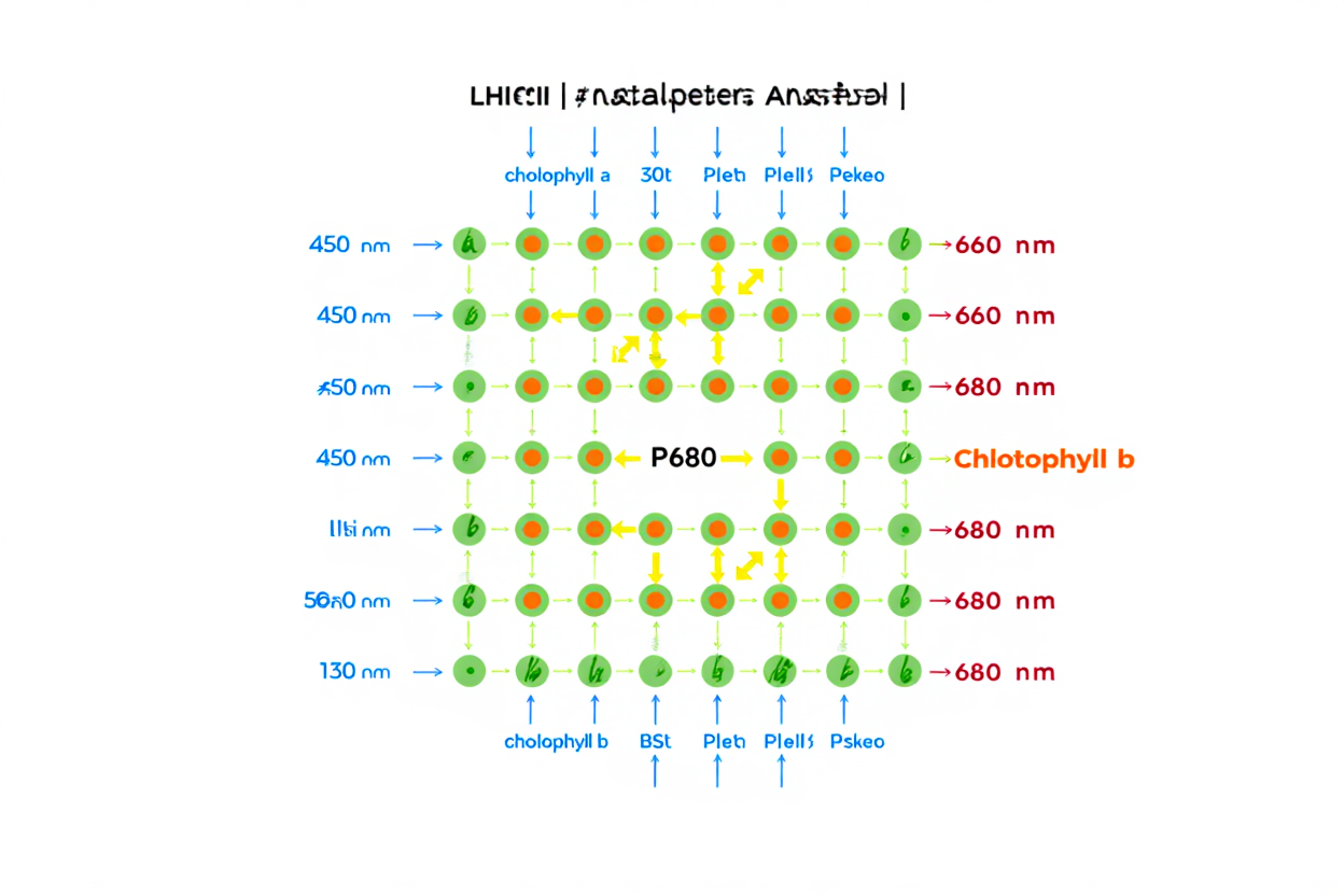
The photosynthetic pigments, such as chlorophylls a and b as well as the carotenoids, form a sophisticated light-capturing system. Organized within the thylakoids into light-harvesting antenna complexes (LHCII), these molecules — nearly 300 chlorophyll molecules per antenna — act like a net to intercept photons and channel them toward the reaction center.
At the heart of each photosystem II lies a special pair of chlorophyll a molecules called P680. It absorbs photons at 680 nm. Excited by this light energy, P680 becomes unstable and quickly gives up an electron, triggering a series of electron transfers. Photosystem I, meanwhile, uses its P700 pair, absorbing at 700 nm, to trigger the reduction of NADP⁺. Together, these two systems create a continuous flow of electrons across the thylakoid membrane.
Water photolysis and the electron transport chain
Once it has lost its electron, oxidized P680 absolutely must replace it. The plant solves this problem by splitting water molecules via an enzymatic complex of photosystem II. Two water molecules are thus broken down during the light phase according to the reaction: 2 H₂O → O₂ + 4 H⁺ + 4 e⁻. This mechanism, called photolysis, releases oxygen, protons (H⁺) into the lumen, and the electrons that regenerate P680.
- Water photolysis: Splitting of H₂O producing O₂, H⁺ and electrons.
- Plastoquinone: The first mobile carrier to receive electrons from photosystem II.
- Cytochrome b6f complex: A proton pump that transfers electrons while reinforcing the gradient.
- Plastocyanin: A soluble carrier that conducts electrons to photosystem I.
After photosystem II, electrons pass through plastoquinone, the cytochrome b6f complex, then plastocyanin before reaching photosystem I. Although their energy decreases with each transfer, this drop is used to actively pump protons into the lumen, creating a gradient used as an energy source.
Stimulated by photons at 700 nm, photosystem I transmits electrons to ferredoxin, then to ferredoxin-NADP⁺ reductase. This enzyme completes the reduction of NADP⁺ into NADPH, an energy-carrying molecule destined for the Calvin cycle in the stroma. Together, this all works like a micro power plant continuously producing ATP and NADPH!
Photophosphorylation and ATP/NADPH synthesis
The accumulation of protons in the thylakoid lumen generates an extremely powerful force. This imbalance forms both a chemical gradient (concentration) and an electrical one (charge), creating a proton-motive force that powers ATP synthase.
ATP synthase is a true molecular turbine embedded in the membrane. When protons flow back from the lumen to the stroma through this enzyme, the energy released allows ATP to be synthesized from ADP. A single unit can produce about a hundred ATP molecules per second, a feat repeated millions of times in every sun-exposed leaf.
| Carrier/Complex | Main function | Location |
| Photosystem II (P680) | Light absorption and water photolysis | Thylakoid membranes of the grana |
| Plastoquinone (PQ) | Electron transport and proton pumping | Thylakoid membrane |
| Cytochrome b6f complex | Electron transfer and increase of the H⁺ gradient | Thylakoid membranes and stromal lamellae |
| Plastocyanin (PC) | Electron transport to PSI | Thylakoid lumen and membrane |
| Photosystem I (P700) | Light absorption and NADP⁺ reduction | Mainly stromal lamellae |
| ATP synthase | ATP synthesis via proton gradient | Thylakoid membrane |
Measurements, for example on lettuce grown under LED light, show oxygen production of about 12 µmol O₂·m⁻²·s⁻¹. This is proof that these photochemical and biochemical reactions are indeed at work in plants. The beauty of the system lies in its efficiency: the ATP and the NADPH produced are immediately used in the stroma to feed the Calvin cycle, with no energy loss.
Calvin cycle and stromal carbon fixation
Once the reserves of ATP and NADPH are built up during the light phase, the plant begins the true synthesis process: sugar-making! This series of reactions, called the Calvin cycle, takes place entirely in the stroma, independently of the thylakoid membranes, using the newly produced energy. Often referred to as the dark phase or light-independent reactions, this stage converts inorganic carbon dioxide into vital glucose, the true foundation of all organic matter production in plants.

Carboxylation of RuBP by Rubisco
The Calvin cycle begins with a decisive step: carbon fixation. The Rubisco enzyme plays an essential role here by catalyzing the binding of a CO₂ molecule to ribulose-1,5-bisphosphate (RuBP), a five-carbon sugar abundant in the stroma. This reaction immediately produces two molecules of 3-phosphoglycerate (3-PG), a three-carbon compound that starts the transformation toward glucose.
- Ribulose-1,5-bisphosphate (RuBP): The CO₂-accepting molecule, continuously regenerated throughout the cycle.
- Rubisco: The most abundant enzyme on Earth, essential to biological carbon fixation.
- 3-phosphoglycerate (3-PG): The direct result of carboxylation, containing the carbon from CO₂.
- Energy requirements: One molecule of ATP is consumed for every full turn of the cycle.
Rubisco isn't just any enzyme: it's truly the most critical protein for life. Without it, no atmospheric carbon could be converted into organic sugar, which would bring photosynthesis to an abrupt halt. For plants grown under intense LED lighting, raising the CO₂ level up to 800 ppm saturates Rubisco and optimizes carbon fixation, boosting leaf sucrose content in just a few hours.
Reduction and regeneration: G3P production
As soon as 3-PG is formed, it undergoes a crucial transformation. ATP from photosynthesis drives the chemical reduction of 3-PG into glyceraldehyde-3-phosphate (G3P), a sugar that stores the energy from carbon dioxide originally captured. This step also requires NADPH, the electron carrier from the photosystem, consuming 3 ATP and 2 NADPH for every CO₂ molecule fixed, which highlights the essential link between the light phase and the dark phase.
- ATP: ATP produced by photophosphorylation activates phosphorylation of 3-PG.
- NADPH: NADPH enables the reduction of 3-PG into energy-rich glyceraldehyde-3-phosphate.
- Glyceraldehyde-3-phosphate (G3P): A reduced sugar serving as the structural basis for all carbohydrates.
- RuBP regeneration: By consuming additional ATP, RuBP is regenerated from G3P.
G3P then becomes a central metabolic intermediate. Part of this G3P leaves the cycle to form glucose, starch or other carbohydrates useful for storage and transport. However, most of it is used to regenerate RuBP through a series of complex reactions, ensuring the cycle continues. To synthesize a single glucose molecule, six molecules of carbon dioxide must be fixed and 18 ATP consumed along with 12 NADPH, all produced thanks to the light phase and light energy.
Every full turn of the cycle releases a water molecule, revealing the overall equation of photosynthesis: 6 CO₂ + 6 H₂O + light energy → 1 glucose + 6 O₂. So-called C3 plants, like wheat, fix CO₂ directly through this mechanism, while C4 plants, such as corn, pre-concentrate it first, which improves their yield in hot environments.
C3 vs C4 differences and the fate of the products
Not all plant species follow the simplified Calvin cycle pattern seen in wheat (C3 plants). C4 plants, adapted to hot, sunny climates, have developed a more efficient strategy: they concentrate carbon dioxide into four-carbon molecules within the mesophyll cells. This carbon is then transported to the bundle-sheath cells to be released directly next to Rubisco, optimizing the efficiency of photosynthesis.
The G3P produced in the chloroplast follows three main paths depending on the plant's needs. A small portion leaves the chloroplast to feed, via the phloem, non-photosynthetic organs such as roots or fruit. Most of the G3P is used to regenerate RuBP to keep the cycle running. Finally, a significant portion is converted into glucose, then polymerized into starch right inside the chloroplast, forming an energy reserve immediately available for the night.
Explanatory diagram and complete educational visualization
Now that you have all the pieces, let's put them together to form a comprehensive, coherent photosynthesis diagram. A good representation turns your grow space into a real laboratory where every component finds its place. Visualizing the chloroplast's organization and connecting the light phase and the dark phase strengthens your understanding, helping you calmly optimize your plants' development.
Structural elements to include in the diagram
To create a truly useful photosynthesis diagram, every major component and its interconnections must be included. Start by drawing the double membrane of the chloroplast, which clearly delimits the stroma inside from the rest of the plant cell. Inside, draw the grana as distinct stacks, ideally colored to contrast with the background.
- Outer and inner envelope: Two clean lines separating the intermembrane space from the surrounding stroma.
- Stroma: An aqueous fluid (shown in blue-green) containing enzymes, DNA, ribosomes and lipid droplets.
- Grana: Vertical stacks of thylakoids (shown in red-orange), often comprising 30 to 50 structures per chloroplast.
- Stromal lamellae: Thin membranes connecting the grana to one another to ensure the continuity of the internal network.
In this stroma, position key elements such as RuBP, starch as it accumulates, and lipid reserves. At the edge of a plant cell, illustrate the entry of carbon dioxide through the stomata before its diffusion toward the stroma. This precise layout helps visualize carbon's complete journey through the complex structure of this organelle.
Annotating flows and photosynthetic processes
The strength of a diagram lies in the arrows that illustrate transformations and energy transfers. Show the path of electrons from PSII to cytochrome b6f, then to PSI, finally reducing NADPH. Also use arrows to show the accumulation of protons that creates the gradient needed for ATP synthesis.
Represent ATP synthase as a small membrane turbine that generates ATP directly in the stroma. Note that PSII is mainly found in the grana, while PSI is more common in the lamellae. These energy flows then feed the Calvin cycle, making chloroplast biology visual and accessible.
Observation methods and hands-on experiments
Want to observe these mechanisms yourself? A simple experiment with Elodea and water enriched with sodium bicarbonate lets you watch oxygen bubbles appear under the effect of light. It's direct proof that water photolysis releases this gas, connecting theory to biological reality.
Under a microscope, you can make out chloroplasts as green granules inside the plant cell. With a drop of Lugol's iodine, the stored starch turns dark, revealing the zones of active carbohydrate production. These concrete observations bring diagrams to life and firmly anchor how photosynthesis works in your memory.